Mapping实验可标测心房、心室组织传导速度
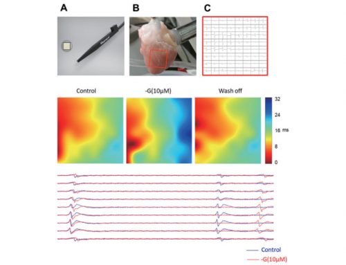
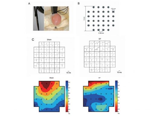
此案例中,科研工作者发现了MKK4基因敲除小鼠心脏Cx43减少,在投稿Circulation后被要求补做mapping实验,从电生理功能上证明Cx43减少影响了左室的动作电位传导速度并因此构成了心律失常的形成基础。如图A,64通道电极放置于小鼠左室,中间电极点给电刺激,可看出基因敲除鼠的AP传导速度明显慢。心室肌传导速度和心肌走向相关,因此文中分析了横向纵向两种传导速度(图B)。
关于心室的mapping实验完成后,作者又标测了左右心房,发现老年基因敲除鼠的心房电传导异常(图C,D),又发表了第二篇论文。

(A). MKK4cko-TAC mice are more susceptible to ventricular arrhythmias and slowed conduction velocity. (B). Most activation maps from MKK4F/F hearts showed a sequential activation pattern, beginning with a localized activation of one region of the matrix followed by its successive spread to the remaining area from which recordings were made. However, the old MKK4CKO hearts often showed a more disordered excitation sequence.
References:
- Zi M, Kimura T E, Liu W, et al. Mitogen-activated Protein Kinase Kinase 4 Deficiency in Cardiomyocytes Causes Connexin 43 Reduction and Couples Hypertrophic Signals to Ventricular Arrhythmogenesis[J]. The Journal of Biological Chemistry, 2011, 286(20): 17821.
- Davies L, Jin J, Shen W, et al. Mkk4 Is a Negative Regulator of the Transforming Growth Factor Beta 1 Signaling Associated With Atrial Remodeling and Arrhythmogenesis With Age[J]. Journal of the American Heart Association: Cardiovascular and Cerebrovascular Disease, 2014, 3(2).


