https://doi.org/10.1186/s12938-020-00763-6
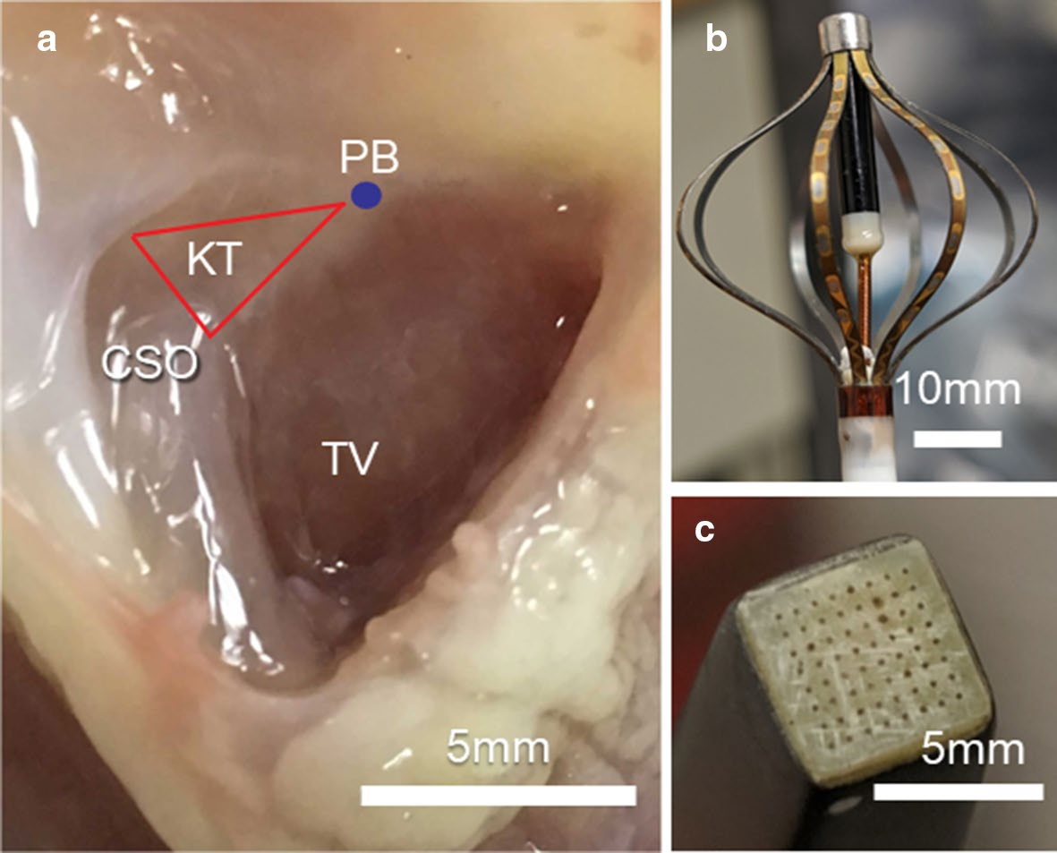
a Right atrial and upper ventricular septum of a perfused rabbit heart. CSO coronary sinus ostium, KT Koch’s triangle, PB penetrating bundle, TV tricuspid valve.
b 64-channel Intellamap Orion basket catheter (Boston Scientifc, Marlborough, MA), which was placed in the LV endocardium.
c 8×8 MappingLab electrode array placed over the region of HB
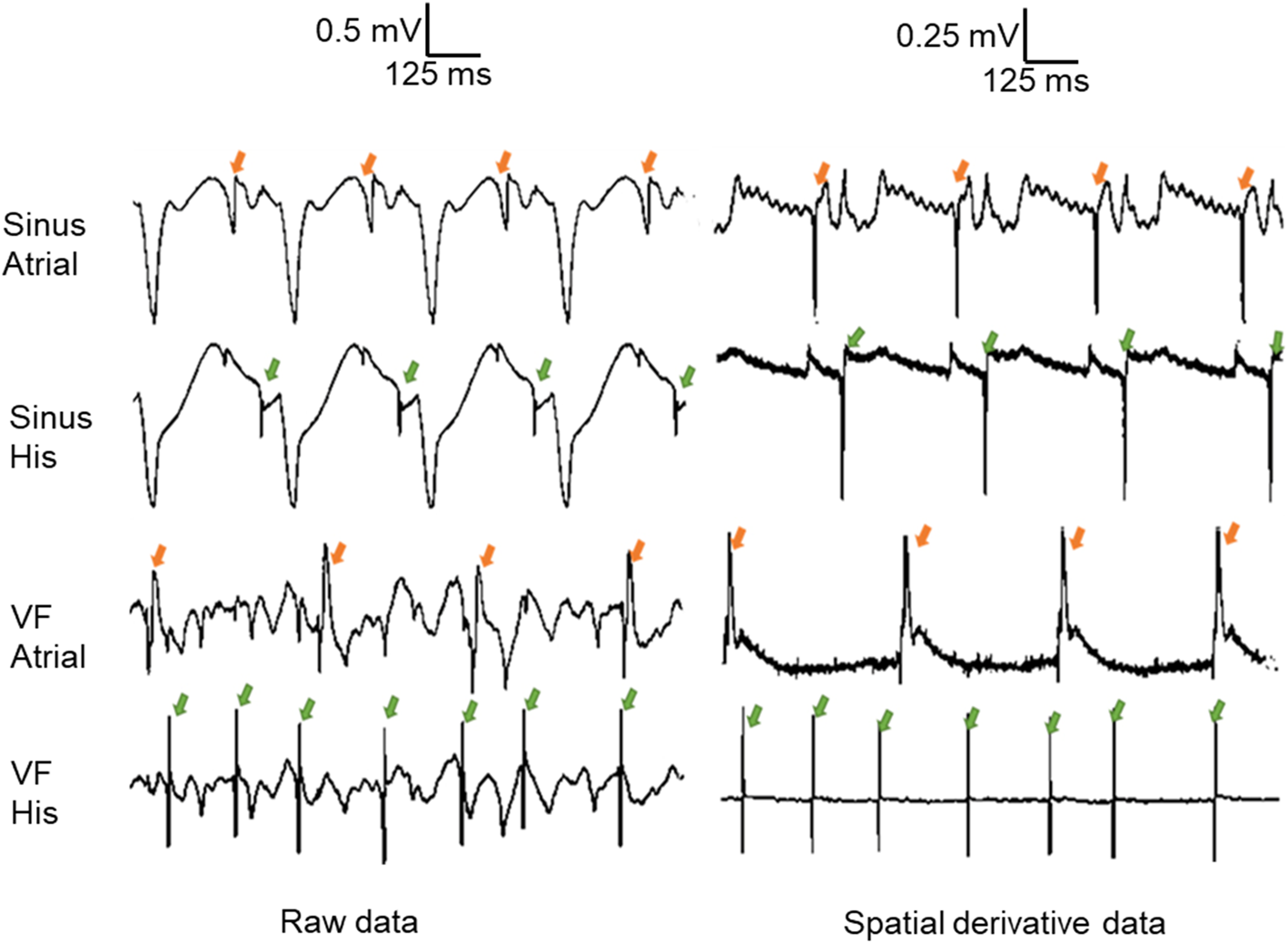
Selected traces with prominent atrial signal and prominent HB signal recorded using the real-time stimulation recording system. Arrows in orange point to atrial activations. Arrows in green point to HB activations. Left column: raw traces during normal sinus rhythm (top two) and VF (bottom two). Right column: spatial derivative traces during normal sinus rhythm (top two) and VF (bottom two). All atrial and HB traces are from the same respective electrodes

